Leg Bones Diagram / Lower Leg Bones Hd Stock Images Shutterstock / Includes obj for maximum compatibility.
Leg Bones Diagram / Lower Leg Bones Hd Stock Images Shutterstock / Includes obj for maximum compatibility.. Your leg bones are the longest and strongest bones in your body. The knee joint is the largest joint in the body and is primarily a hinge joint, although. The foot bones shown in this diagram are the talus, navicular, cuneiform, cuboid, metatarsals and calcaneus. Quizzes on human skeletal system anatomy, bone anatomy, and bone markings. Health diagram bone skeleton leg knee science anchor chart human human body.
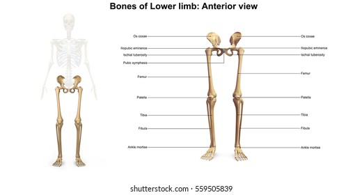
The femur, or thigh bone, is the largest, heaviest, and strongest bone in the human body. At the distal end of the femur, two rounded condyles meet the tibia and fibula bones of the lower leg to form the knee joint. The femur, or thighbone, is the longest and largest bone in the human body. The knee is a strong but flexible hinge joint. File is ready to render.
Diagram Body Bone Diagram Full Version Hd Quality Bone Diagram Ritualdiagrams Partytimebanqueting It from image.shutterstock.com Want to learn more about it? Lower jaw (mandible) collar bone. Quizzes on human skeletal system anatomy, bone anatomy, and bone markings. They are primarily compact bone but may have a large amount of spongy bone at the ends or extremities. Learn vocabulary, terms and more with flashcards, games and other study tools. Learn how to draw the femur, patella, tibia, and fibula in this lesson! The humerus and the femur are corresponding bones of the arms and legs, respectively. The bone that goes from your pelvis to your knee is called the femur (say:
The bones of your leg have roughened patches on their surfaces where muscles are attached.
At the distal end of the femur, two rounded condyles meet the tibia and fibula bones of the lower leg to form the knee joint. Download the free graphic resources in the form of png, eps, ai or psd. The femur, or thigh bone, is the largest, heaviest, and strongest bone in the human body. The tibia is the main bone of the leg, forming what is more commonly known as the shin. Learn vocabulary, terms and more with flashcards, games and other study tools. Lower jaw (mandible) collar bone. The foot bones shown in this diagram are the talus, navicular, cuneiform, cuboid, metatarsals and calcaneus. When your muscles contract, they pull the bone they're. Visit kenhub for more skeletal system quizzes. The foot bones shown in this diagram are the talus, navicular, cuneiform, cuboid, metatarsals and calcaneus. Skeleton leg ankle joints and toe phalanges, cuboid, metatarsal, navicular and cuneiform bones, hand drawn dorsal view of foot. The information above used to be on the page 'skeletal structures of the feet and hands' in the form of simple labelled diagrams of the leg. The foot bones shown in this diagram are the talus, navicular, cuneiform, cuboid, metatarsals and calcaneus.
Pngtree offers bone diagram png and vector images, as well as transparant background bone diagram clipart images and psd files. When your muscles contract, they pull the bone they're. Learn how to draw the femur, patella, tibia, and fibula in this lesson! Your leg bones are very large and strong to help support the weight of your body. Includes leg (femur, tibia, patella, and fibula) and foot (tarsals and digits) bones.
Leg And Knee Anatomy Bones Muscles Soft Tissues Kenhub from thumbor.kenhub.com The knee joint is the largest joint in the body and is primarily a hinge the bones of the leg are the femur, tibia, fibula and patella.the foot bones shown in this diagram are the talus, navicular, cuneiform, cuboid. At the distal end of the femur, two rounded condyles meet the tibia and fibula bones of the lower leg to form the knee joint. The foot bones shown in this diagram are the talus, navicular, cuneiform, cuboid, metatarsals and calcaneus. Learn how to draw the femur, patella, tibia, and fibula in this lesson! Time to jump right into the biggest and strongest bones in the human body. The femur, or thigh bone, is the largest, heaviest, and strongest bone in the human body. Its lower end helps create the knee joint. Most bones (particularly the long bones of the arms and legs — which make up the appendicular skeleton) have a hard outer shell known as cortical bone.
Health diagram bone skeleton leg knee science anchor chart human human body.
You'll learn about the muscles, bones, and other structures of each area of the leg. Arm bones (= bones in arm) are part of the appendicular skeleton which includes the hands, arms and shoulder girdle (clavicle and scapula) and the feet note: Human foot bones anatomy sketch of orthopedics medicine. They are primarily compact bone but may have a large amount of spongy bone at the ends or extremities. The foot bones shown in this diagram are the talus, navicular, cuneiform, cuboid, metatarsals and calcaneus. Continue scrolling to read more below. High resolution textures and displacement included. At the microscopic level, this hard outer shell is made up of rod like structures called osteons. Long bones include bones of the thigh, leg, arm, and forearm. At the distal end of the femur, two rounded condyles meet the tibia and fibula bones of the lower leg to form the knee joint. Its lower end helps create the knee joint. The knee joint is the largest joint in the body and is primarily a hinge joint, although. The femur, or thighbone, is the longest and largest bone in the human body.
The foot bones shown in this diagram are the talus, navicular, cuneiform, cuboid, metatarsals and calcaneus. Includes leg (femur, tibia, patella, and fibula) and foot (tarsals and digits) bones. The tibia is the main bone of the leg, forming what is more commonly known as the shin. Master leg and knee anatomy using our topic page. The femur, or thighbone, is the longest and largest bone in the human body.
Lower Leg Bones Hd Stock Images Shutterstock from image.shutterstock.com They allow you to move and provide support for your upper body. Includes leg (femur, tibia, patella, and fibula) and foot (tarsals and digits) bones. Long bones include bones of the thigh, leg, arm, and forearm. Its lower end helps create the knee joint. File is ready to render. Learn how to draw the femur, patella, tibia, and fibula in this lesson! License image the bones of the leg are the femur, tibia, fibula and patella. Includes obj for maximum compatibility.
The information above used to be on the page 'skeletal structures of the feet and hands' in the form of simple labelled diagrams of the leg.
You'll learn about the muscles, bones, and other structures of each area of the leg. Your legs are two of your most important body parts. It mainly serves as an attachment point for the muscles of the lower leg. The knee joint is the largest joint in the body and is primarily a hinge joint, although some sliding and rotation occur. Includes obj for maximum compatibility. Time to jump right into the biggest and strongest bones in the human body. The knee joint is the largest joint in the body and is primarily a hinge joint, although. Human leg bones vector image. The femur, or thigh bone, is the largest, heaviest, and strongest bone in the human body. Visit kenhub for more skeletal system quizzes. At the distal end of the femur, two rounded condyles meet the tibia and fibula bones of the lower leg to form the knee joint. Your leg bones are very large and strong to help support the weight of your body. Includes leg (femur, tibia, patella, and fibula) and foot (tarsals and digits) bones.


Comments
Post a Comment